
By examining the dissolved movement such as potassium yoisid and glucose, and tracking the changes in weight in the various sugars concentrations, the experiment aims to show the principles of passive transfer and how the focus gradients affect the molecular movement through the membranes.
This experiment is looking at how changing solvent concentrations affect the osmosis and spreads through a semi -science membrane (dialysis tubes), studying the movement of water, dissolved, and the resulting changes in the properties of the solution and weight over time.
introduction
The osmosis and spread of the basic processes of cellular transport, and play important roles in maintaining balance and enabling basic biochemical functions (Alberts et al., 2014). Both forms of negative transport, allowing molecules to move through biological membranes without energy. The osteoporosis is defined as the movement of water molecules through a semi -capable membrane to carry out a less concentration area to a higher concentration area until the balance is reached. On the other hand, the spread of dissolved molecules involves a higher concentration area to a lower concentration area (Binod, 2024).
Understanding the dynamics of these processes is necessary to explore how cells interact with their environment, absorb nutrients and eliminate waste. There are three main types of solutions when it comes to osmosis: excessive tension, where there are more solvents outside the cell, which causes water to flow out of the cell; Hypotonic, where there is less melted outside the cell, which leads to the transmission of water to the cell; And isotonic, where the concentration of dissolved is the same inside and outside the cell, which leads to the lack of a clear water movement (Binod, 2024).
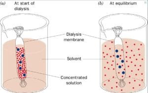
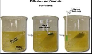
In this experiment, dialysis bags are used in the experiment because they are deficient and represent the artificial cells to study osmosis and spread. The hypothesis of the experiment is that the starch/glucose solution inside the dialysis tubes will show a change in color due to the spread of potassium yolis. The weight of dialysis bags will change due to the osmosis, depending on the concentrations of scrubs.
Materials and methods
Steps to study spread and osmosis using dialysis tubes
Experience:
- Prepare a spread solution:
- Fill a small dorm with water.
- Add 10 drops from the potassium yoideen solution to the beacon to achieve a medium brown color.
- Watch for proliferation:
- Record the initial color and the appearance of potassium yoisid solution in the beacon.
- Wait for the solution to publish, with a color change with the solution until the solution becomes yellow all the time, indicating complete spread.
- Preparing dialysis tubes:
- Cut a piece of dialysis tubes about 10 cm.
- Connect one end of the tubes safely and leave the other end open.
- Fill in dialysis tubes:
- Fill the open end of dialysis pipes about two -thirds full of starch/glucose solution.
- Safely connect the open tip for the tubes.
- Initial notes record:
- The color of both the potassium yoideen solution and the starch/glucose solution in the dialysis tubes in Table 1.
- Lower the glucose test tape in the ring solution and record the result in Table 2.
- Put the dialysis tubes in the beach:
- Diplomatic dialysis tubes in the beads that contain potassium yoisien solution.
- Wait and monitor:
- After 30 minutes, register color changes in both potassium yoisal solution and starch/glucose solution inside dialysis tubes in Table 1.
- Cover the glucose test tape in the ring solution again and record the result in Table 2.
Epiprus experience:
After 60 minutes, record the final weights of all dialysis cells in Table 3.
Preparing osmosis solutions:
- Fill a small dormation of about two -thirds full of Skrouz solution by 25 %.
- Fill a large dormor of about two -thirds of the scrub solution by 1 %.
Preparing dialysis pipes for artificial cells:
- Get four pieces of soaked dialysis tubes.
- Cut one end of each piece of dialysis tubes with a clip. Naming clips A, B, C and D.
Fill in dialysis tubes:
- Open “Cell A” and fill it about two -thirds of a full of sutures, and then remove it safely.
- Fill the “cell B” with a 1 % sugar solution, “Cell C” with a 10 % sugar solution, and “Cell D” with sugar solution by 25 %, and they discourage each of them.
Initial weights record:
- The weight of each of the four dialysis cells and the registration of their initial weights in Table 3.
Dialysis cells:
- Put the “Cell A” in the small across with the Skuzuz solution by 25 %.
- Put the “cell B, C and D” in the large acid with the Skros solution by 1 %.
Wait for weight:
- After 15 minutes, remove the cells from their cups, dry them a little, and weigh them.
- Record the new weight in Table 3.
- Return the cells to their cups.
Repeat the weight process:
- Repeat the process of removing, drying, weighing weight after 30 minutes, 45 minutes and 60 minutes.
Final weights:
- After 60 minutes, record the final weights of all dialysis cells in Table 3.
results
The results of the experiment explain the spread of potassium yoisid and glucose behavior in dialysis tubes, as shown in the following tables.
Table 1. Potassium iodine solution spreads from the beacon to starch/glucose solution from dialysis tubes
| Potassium yulid solution | Starch solution/glucose | |
| The beginning of color | Yellow | white |
| Finish | clear | Purple/cloudy |
Table 2. Glucose tape test for spread
| color | The present glucose? | ||||
| Glucose test bar at the beginning | Wild ducks | negative | |||
| Glucose test bar at the end | Brown/green | Glucose effect | |||
The results of the osmosis experience are clarified through the movement of dissolved through the dialysis tubes and the changes in weight, indicating the nature of the environment, hyperthyroidism, or medium tension, or equal tension.
Table 3. Change in the weight of dialysis cells as a time function
| 0 minutes (initial weight) (GM) | 10 minutes weight (GM) | Weight 20 minutes (GM) | 30 minutes weight (GM) | 40 minutes weight (GM) | |
| Cell a | 24.16 | 24.53 | 23.90 | 24.12 | 24.65 |
| Cell b | 19.27 | 19.31 | 19.34 | 19.30 | 19.33 |
| Cell c | 24.67 | 24.58 | 24.33 | 24.01 | 23.79 |
| Cell d | 28.50 | 26.87 | 25.65 | 25.57 | 24.78 |
discussion
The experiment showed that the potassium yoisin spreads in the starch/glucose solution inside the dialysis tubes, causing the color change and confirmation of successful spread. In addition, the weight of dialysis bags varied based on the surrounding sugar concentrations, indicating the effects of the osmosis: cells were lost in excessive weight solutions, while those in the solutions are equal tension remained stable.
Initial observations revealed that the potassium yoideen solution started as a yellowish brown, while the starch/glucose solution was white. After 30 minutes, the potassium yoideen solution
It has become clear, and the starch solution/purple glucose/cloudy turns, indicating a successful spread of iodine in the tubes.
The glucose test tape initially showed the color of the wild ducks, indicating the absence of glucose. At the end of the experiment, the tape has changed to brown/green, which confirms the effect of glucose in the acurnum solution.
Weight changes over time indicate varying responses to the surrounding solutions. The cell A showed a slight increase in weight after 10 minutes, but then fluctuating indicates an excessive tension environment in the beach. The B cell remained relatively stable indicating an equal tension environment. On the other hand, the C cell showed a gradual decrease in weight, while the cell D showed a significant decrease, reflecting the water loss due to its excessive environment. The dissolved is transmitted from the highest focus to the low focus to maintain balance (Alberts et al., 2014).
The experiment had weaknesses, such as the differences in the extent to which dialysis pipes allowed the material to pass, which may lead to uneven results. Also, the lack of control of temperature and dependence on color changes to measurements may make the results less reliable.
In general, the hypothesis results support that the spread occurs through the dialysis membrane and highlights the effects of stress on the weight of dialysis bags.
Other experiences
Future experiments can be studied in how the temperature affects osmosis and spread, with the expectation that high temperatures make these processes occur faster. We can also try different materials, such as salt or sugar, to see how the way the molecules move change.
Reference
Alberts, B., Johnson, A., Lewis, J., RAFF, M., Roberts, K., & Walter, P. (2014). The molecular biology of the cell (Sixth Edition). Wreath Science.
GC, Binod. “Dhumination and spread: the differences and factors that affect them.” Note science14 April 2023. October 2, 2024. Osmosis and spread: the differences and factors that affect them
Mika, TA, Klein, RJ, Bullerjahn, AE, Connour, RL, Swimmer, LM, White, Re,
Gosses, MW, Carter, TE, Andrews, AM, Maier, JL, & Sidiq, F. (EDS.). (2024). Anatomy and biotechnology 211 laboratory guide (Third Edition). Owens Community College.
GC, Binod. Cellular transport: negative and active mechanisms. Note scienceSeptember 3, 2024, October 2, 2024. Cellular Transport: Negative and Activated Mechanisms – Scientific Notes